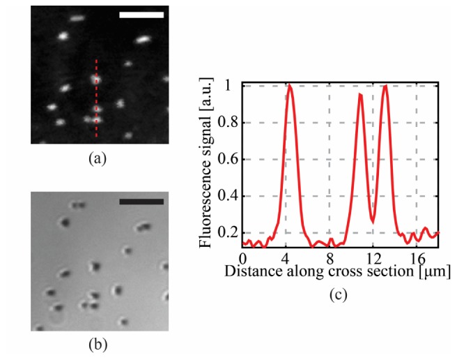
Fig. 5.

Demonstration of high resolution fluorescence imaging through the CWG: (a) Fluorescence image of 1.5 μm beads in a plane 100 μm away from the CWG facet. The sample is placed in front of the silica part of the CWG in order to maximize resolution and signal collection. The FWHM of the spot is 1.5 μm and the scanning step is 0.31 μm; (b) white light optical image of the sample; (c) cross-sectional plot along the red dashed line in (a) shows that two beads 2.2 μm far apart were completely resolved, giving an upper limit for the resolution of the fluorescence endoscope of about 2 μm. Scale bar are 10 μm.
