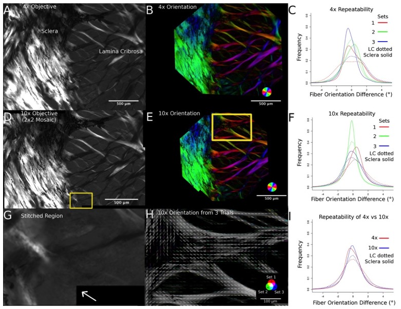
Fig. 7.
Repeatability in optic nerve head tissue. Three image sets were captured using 4x and 10x objectives of a section of sheep optic nerve head tissue (A and D) and for each image set, the fiber orientation was calculated (B and E). The measurements were highly repeatable regardless of resolution (I) and tissue density (C and F) and there were no stitching artifacts (arrow in G indicates stitched region). The frequencies in panels C, F and I have been normalized to the total number of measurements. The fiber orientations from each image set were color coded and compared such that white indicated perfect agreement, and colors represented differences. The majority of the orientation lines are white (H).

