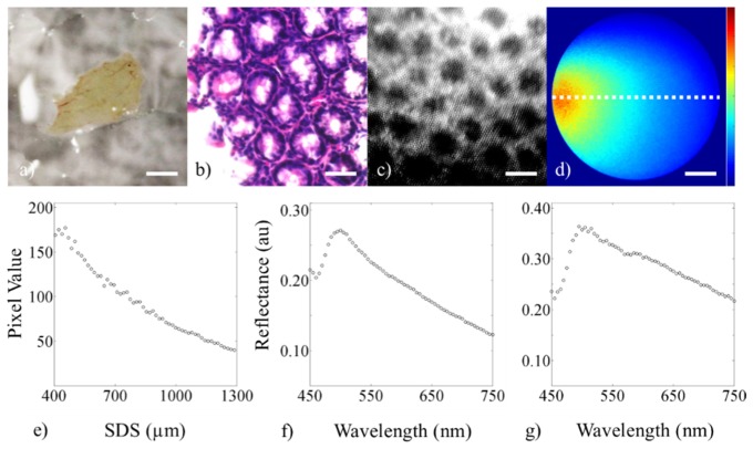
Fig. 7.
Demonstration of the three modalities showing data from a 16-week old wild-type (C57BL/6J) male mouse. The figure shows (a) digital image of the 4-5 cm colon tissue (lumen side facing up, scale bar = 5 mm), (b) histology of an adjacent section (scale bar = 50 µm), (c) cropped and enhanced high-resolution fluorescence image after topical staining with 0.01% w/v proflavine (scale bar = 50 µm), (d) sDRIM data (scale bar = 225 µm, color bar = 0-200), (e) quantification of the sDRIM data taken across the face of the image fiber (400-1,300 µm SDS from 635 nm laser source), (f) broadband sDRS data (374 µm SDS), and (g) broadband sDRS data (730 µm SDS).

