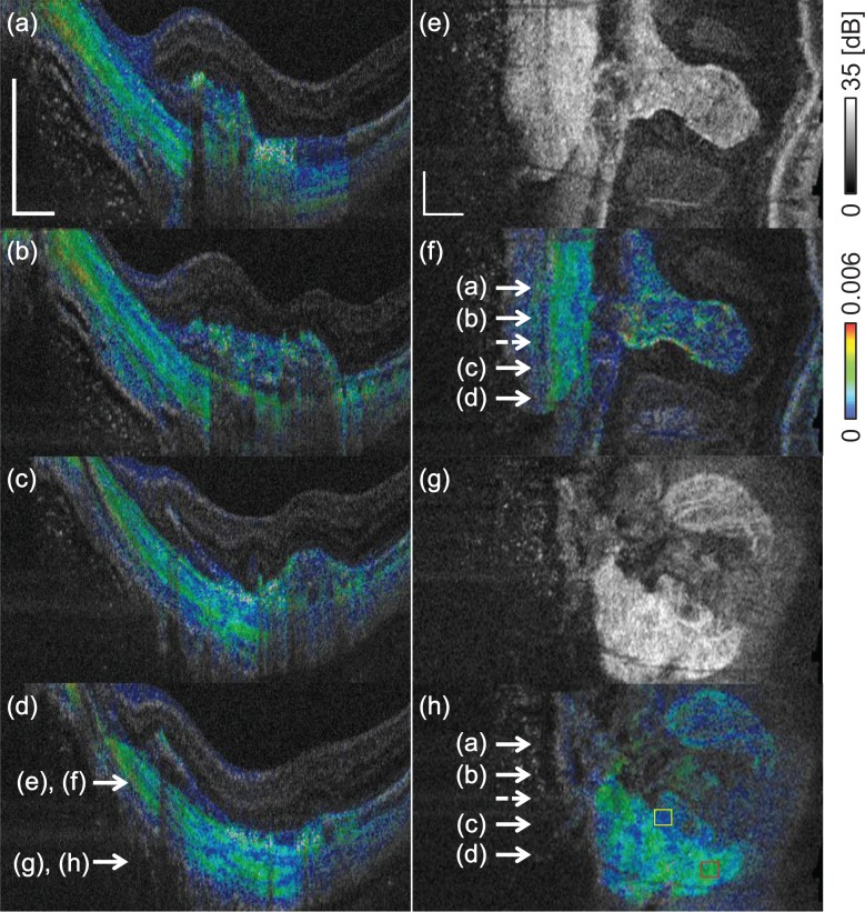
Fig. 9.
Birefringence cross-sections (a)–(d) show the layered birefringence structure of the sclera. (e) and (g) show en face scattering OCT slices, and (f) and (h) are the corresponding birefringence slices, respectively. The transverse positions of the cross-sectional images are indicated by the solid arrows in (f) and (h). The dashed arrow indicates the position corresponding to Figs. 8(b)–(e). The depth positions of the en face images are indicated by the arrows in (d). The scale bar represents 0.5 mm × 0.5 mm.

