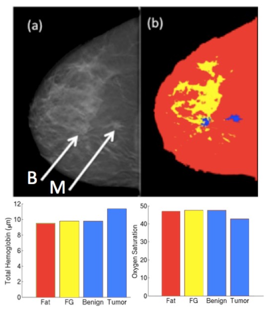
Fig. 7.

(a) DBT slice for a patient with benign (left arrow) and malignant (right arrow) lesions. (b) DBT image stack was segmented into adipose, fibroglandular (FG) and two ROIs. (c) Total hemoglobin concentration and oxygen saturation obtained from the NIRST/DBT recovery for each region.
