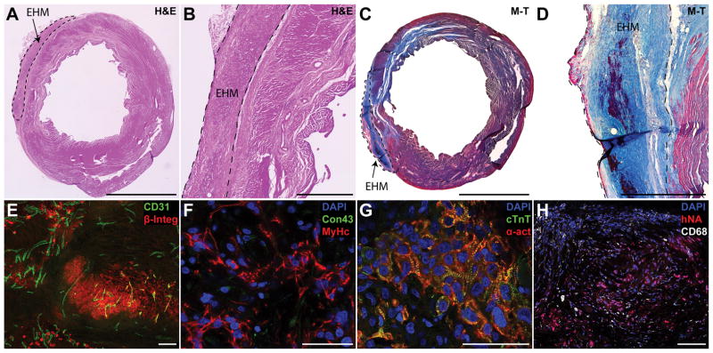
Figure 3. Sizable grafts of human cardiomyocytes could be found one month after EHM implantation.
(A–D) One month after implantation EHM, grafts were found covering the scar area or border zone on H&E or Masson’s trichrome (M–T) sections. EHMs were typically separated from the host by a small layer of fibrotic tissue. (E) Grafts staining positive for human beta integrin 1 (β-Integ) could be found for all EHM hearts assessed. Grafts were perfused by host vessels (CD31) that grew into the graft (maximum intensity projection of a 30 μm section). (F) Grafts consisted primarily of human CMs staining positive for beta myosin heavy chain (MyHc), but there were few cell junctions that stained positive for connexin 43 (Con43). (G) While clearly striated sarcomeres could be found (cardiac troponin T, cTnT; alpha sarcomeric actin, α-act) there were few signs of myofibril alignment. (H) EHMs showed few infiltrating macrophages (CD68) indicating stable grafts. Scale bars A,C: 5 mm; B,D: 1 mm; E,H: 100 μm; F,G: 50 μm.

