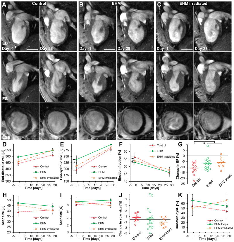
Figure 4. EHM implantation reduces dilative remodelling but this effect is not due to human CM engraftment.
(A–C) The top row shows representative 2-chamber long-axis views at end-diastole (ED) one day prior to and 28 days after EHM implantation or sham surgery (control group). The middle row shows the same hearts at end-systole (ES) while the bottom row shows corresponding late gadolinium enhancement images (LGE) at mid-ventricular level. (D,E) End-diastolic volumes increased in all groups but were not significantly different (P=0.35). End-systolic volumes also increased in all groups, but the control group mas most pronounced compared to all other groups (control n=12 vs. EHM n=14; P=0.05). (F,G) Ejection fractions declined in all groups. The control group showed the highest relative decline compared to EHM implantation groups (P=0.03); however, there was no difference between EHMs or irradiated EHMs that did not contain viable CMs (P=0.19), indicating that beneficial effects on remodelling may not be directly mediated by grafted cells. (H,I,J) No significant difference was observed for changes in scar size over time (P=0.32). (K) There was a trend toward increase in diastolic dysfunction (E′/A′ <1) for hearts receiving irradiated EHM loops while a small decline was observed for the other groups; however, this did not reach statistical significance (P=0.12). Scale bars A–C: 5 mm.

