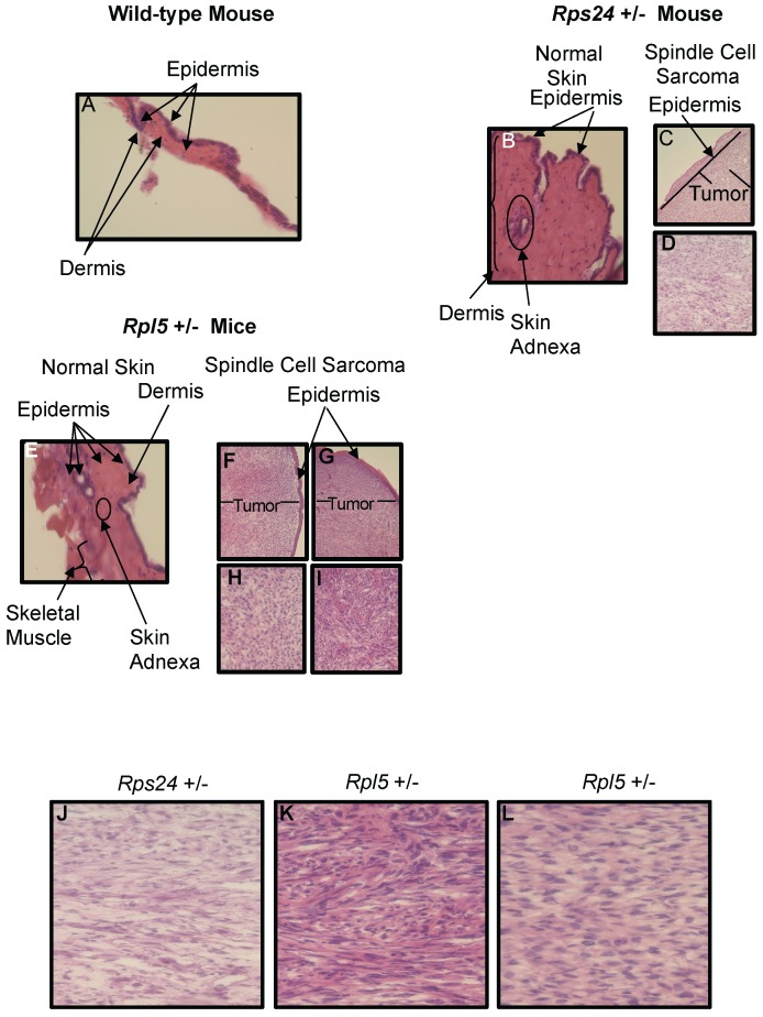
Figure 1.
Histology of Wild-type, Rps24+/-, and Rpl5+/- Normal Skin and Spindle Cell Sarcoma Tissues. Hematoxylin and Eosin staining showed normal epidermis of wild-type (A), Rps24+/- (B), and Rpl5+/- (E). However, there was a uniform localization of spindle tumor cells beneath the epidermis from Rps24+/- mouse (C and D) and Rpl5+/- mice (F, H, G, and I) with Rps24+/- tumor cells (Figure S3 and J) and tumor cells from Rpl5+/- with smaller tumor (Figure S3 and K) having very similar morphological appearances. In contrast, tumor cells from the Rpl5+/- mouse with a larger tumor (Figure S3and L) were more rounded and had clear vacuoles, lesser degree of fascicular architecture and nuclear pleomorphism. All images are at 40X magnification.

