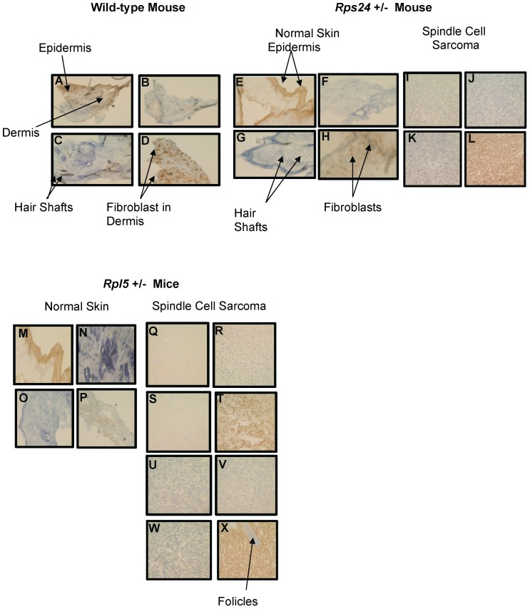
Figure 2.
Immunohistochemical Comparison of Wild-type, Rps24+/-, and Rpl5+/- Normal Skin with Tumor Tissues. Pan-keratin staining was detected throughout the epidermis with no detectable staining in the dermis of wild-type (A), Rps24+/- (E), and Rpl5+/- (M) skin sections, and was also negative in all the tumor tissues (I, Q, and U). Negative staining for both LCA (B, F, J, N, R, V) and S100 (C, G, K, O, S, W) was observed in the dermis of all tissue sections. Vimentin staining throughout the dermis in wild-type (D) and Rps24+/- (H) normal skin tissues corresponded to fibroblasts. In contrast, all the tumor tissues stained very strongly for vimentin (L, T, and X). All images are taken at 40X magnification.

