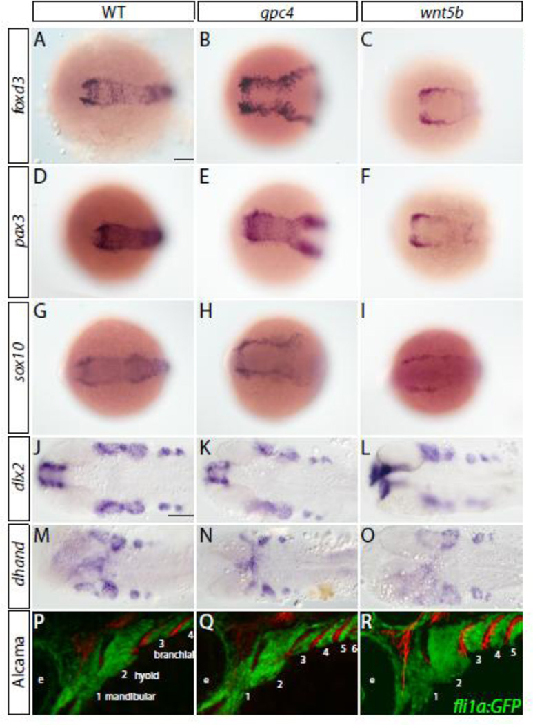
Fig. 3. Cranial neural crest cell migration and specification is unaffected in gpc4 and wnt5b mutants.
(A–O) Neural crest specification appears normal within gpc4 and wnt5b mutants Scale bar = 100 µm. (A– I) Pre-migratory neural crest markers. 6 somites. (J–O) Pharyngeal arch markers. (J–L) 28 hpf. (M–O) 32 hpf. (P–R) Migration of neural crest appears to be unaffected in gpc4 and wnt5b mutants when compared to wild type fish. Tg(fli1a:EGFP)y1 embryos have neural crest derived cells labeled by GFP expression. Embryos were fixed at 33 hpf and stained with the Alcama (Zn5) antibody (red). Pharyngeal arches are numbered. Scale bar = 50 µm.

