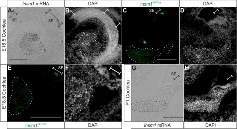
Fig. 5. Insm1 expression subsides in SpG neurons as they differentiate and is undetectable at birth.

Representative sections demonstrating Insm1 expression at E16.5-P1 in the cochlea. (A) In situ hybridization with an Insm1 antisense probe on a coronal section of E16.5 head, which results in an axial (perpendicular to the modiolus) section of the cochlea. Insm1 signal appears reduced in the SpG when compared with E14.5 (Fig. 4A). Insm1 mRNA is also expressed in the sensory epithelium. (B) Nuclear pattern corresponding to (A). (C) Immunohistochemistry for GFP (green) representing Insm1GFP.Cre expression in the E16.5 cochlea. Insm1GFP.Cre signal is decreased in SpG neurons when compared to Fig. 4C, and is also detected in sensory epithelium. (D) Nuclear pattern corresponding to (C). (E) Immunohistochemistry for GFP (green) representing Insm1GFP.Cre expression in modiolar section of E18.5 cochlea. Insm1GFP.Cre signal is further decreased in SpG neurons when compared to previous stages and epithelial pattern appears to correspond to OHCs. (F) Nuclear pattern corresponding to panel (E). (G) In situ hybridization with an Insm1 antisense probe on a modiolar section of P1 cochlea (mid-basal turn) showing no Insm1 mRNA expression. (H) Nuclear pattern corresponding to panel (G). Dotted lines in all panels delineate SpG. Brackets designate sensory epithelium. Scale bar: 200 μm. SpG: spiral ganglion, SE: sensory epithelium.
