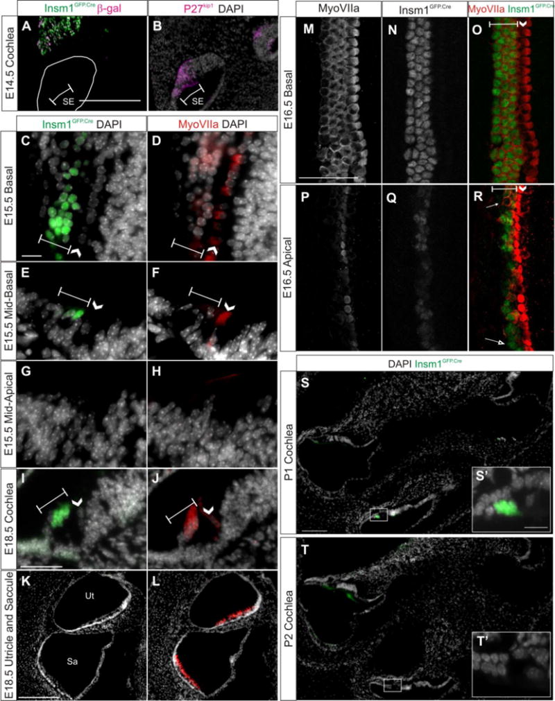
Fig. 6. Insm1 is transiently expressed by nascent OHCs, but not IHCs.

(A,C,E,G,I,K,S,T) Representative sections demonstrating Insm1 expression by immunohistochemistry for GFP (green) in cochlear sections of Insm1GFP.Cre mice at E14.5–E18.5. In (A), immunohistochemistry for lnsm1 lineage marker β-gal is represented in magenta. (A,B) At E14.5 Insm1 expressing cells or their descendants are not in the sensory epithelium (A), which is identified in an adjacent section by immunohistochemistry for P27kip1 (B, magenta). (D,F,H,J,L) Immunohistochemistry for HC marker MyoVIIa (red) in sections adjacent to C,E,G,O,Q, respectively, with nuclear pattern (white). (C,D) At E15.5 in the most basal aspect of the organ of Corti (section provides a glancing view and hence multiple hair cells per row), Insm1GFP.Cre and MyoVIIa are expressed in all rows of OHCs. (E,F) At E15.5 in the mid-basal turn of the organ of Corti, Insm1GFP.Cre is expressed in 2–3 of OHCs and MyoVIIa is only expressed in the first OHC. (G,H) At E15.5 in the mid-apical turn of the organ of Corti, Insm1GFP.Cre and MyoVIIa are not detectable. (M,N,O) Immunohistochemistry for MyoVIIa (M, white), Insm1GFP.Cre (N, white), and the merged image (O, MyoVIIa in red and Insm1GFP.Cre in green) in a whole mount show that, at E16.5 in the basal organ of Corti, Insm1 is expressed in all OHCs. (P, Q, R) Immunohistochemistry for MyoVIIa (P, white), Insm1GFP.Cre (Q, white), and the merged image (R, MyoVIIa in red and Insm1GFP.Cre in green) in a whole mount show that, at E16.5 in the apical organ of Corti, Insm1 is typically expressed in OHCs that are expressing MyoVIIa, with some exceptions where OHCs express MyoVIIa without Insm1 (filled arrow) or vise versa (open arrow). (I,J) At E18.5 throughout the cochlea, Insm1GFP.Cre and MyoVIIa are expressed in all three rows of OHCs, and Insm1 is clearly not expressed in IHCs. (K,L) At E18.5 in the utricle and saccule, Insm1GFP.Cre is not detectable in MyoVIIa expressing HCs. (S–T) Insm1GFP.Cre (green) in sections through two turns of the cochlea at P1 (S) and P2 (T) demonstrating that Insm1 expression persists in apical OHCs through P1 and has mostly subsided throughout the cochlea by P2. (S′ and T′) represent boxed regions in (S and T), respectively. Brackets designate rows of OHCs. Arrow heads indicate the single row of IHCs. Scale bar: (A,Q) 200 μm, (C, I) 50 μm. SE: sensory epithelium, Sa: saccule, Ut: utricle.
