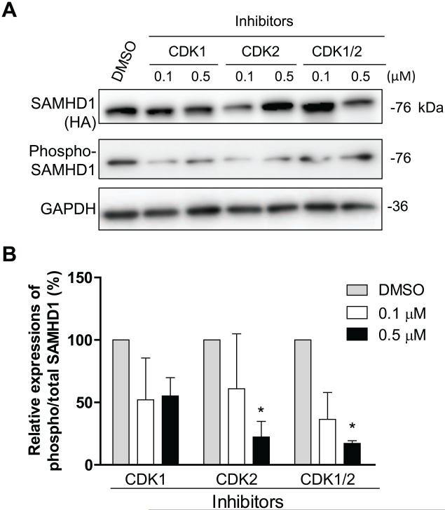
Fig. 4.
Endogenous CDK1 and CDK2 phosphorylate mSAMHD1 at T634 in mouse fibroblast cells. (A) NIH3T3 cells stably expressing mSAMHD1 were treated with inhibitors specific to CDK1 or CDK2 at the indicated concentrations. The effects of each inhibitor on the phosphorylation of mSAMHD1 at T634 were assessed at 24 h post-transfection by immunoblotting with a phospho-specific antibody. (B) An average of three independent experiments. The protein levels were quantified based on the band densities. The levels of total mSAMHD1 and phospho-SAMHD1 were normalized to GAPDH, and then the ratio of phospho/total mSAMHD1 was calculated and the ratio of the vector group was set as 100%. Results are shown as mean ± SD (n=3). The data was analyzed by one-way ANOVA with Dunnett’s test (*, P < 0.05).

