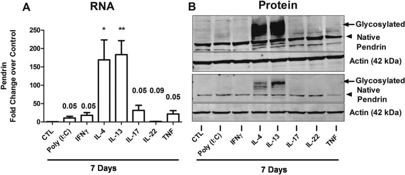
Fig. 5. Pendrin is induced by Th2 cytokines and IL-17A in airway epithelial cells in vitro.
(A) Differentiated nasal epithelial cells (NECs) were untreated (CTL) or stimulated with Toll-like receptor 3 ligand (Poly (I:C)) or cytokines as indicated for 7 days. Pendrin gene expression was quantified using real-time PCR (n=9). Gene expression was normalized to actin and expressed as fold change over untreated control samples. Pendrin protein was verified using immunoblot analysis in stimulated ALI-cultured NECs. Arrow head and arrow indicates native (80 kDa) and glycosylated (100 kDa) pendrin respectively. Shown are representative blots for pendrin and actin from two donors out of 8 donors.

