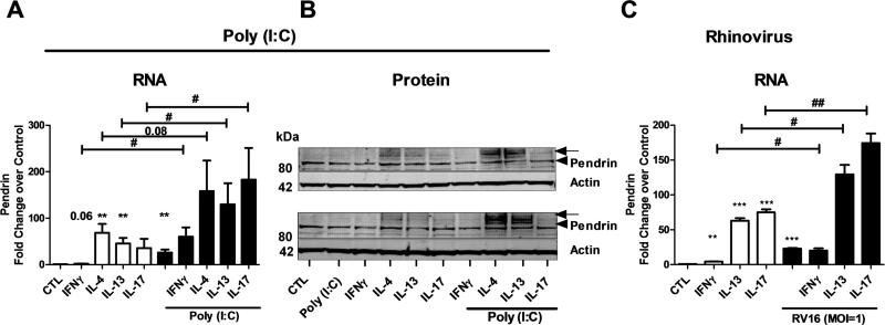
Fig. 9. Potentiation effect of IL-13/IL-17A and Poly(I:C)/ Human Rhinovirus infection on pendrin expression in airway epithelial cells.
Differentiated nasal epithelial cells were stimulated with cytokines and/ or Poly(I:C) (A and B) or cytokines and/or human rhinovirus infection (C). Cells were harvested at 24 h (A) or 48 h (B and C) after stimulation. RNA (A and C) was analyzed using real-time PCR. Pendrin mRNA was normalized to actin and expressed as fold change over control. Data from 8-12 experiments (A); 2 experiments (B) and 4 experiments (C) are shown. B (2) and C (4) experiments. In B, the arrow head and the arrow indicate native (80 kDa) and glycosylated (100 kDa) pendrin respectively. #; *p<0.05, ##; **p<0.01by paired t-test, * comparing control to treated.

