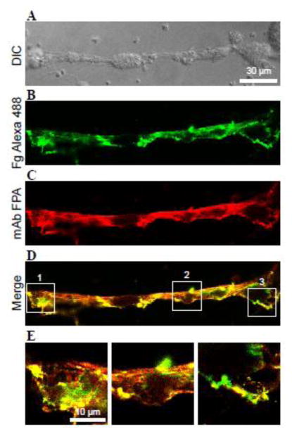
Figure 4. Detection of intact fibrinogen on the surface of thrombi.
Thrombi were generated by perfusing whole blood containing 6 μM argatroban over collagen-coated surfaces for 15 min at high shear rate of 3000 s−1. Thrombi were allowed to rest in Tyrode buffer for an additional 15 min to convert surfaceassociated fibrinogen into fibrin. Subsequently, Alexa 488-labeled fibrinogen in Tyrode buffer containing 100 μM PPACK was perfused at 1500 s−1 for 15 min. A, a representative DIC image of thrombi and connecting fibrin fibers. B, thrombi were perfused with 20 μg/mL fibrinogen containing 1:100 Alexa 488-labeled fibrinogen (green). C, thrombi were subsequently stained with mAb FPA (red). D, fluorescent images of fibrinogen and fibrin were merged to highlight colocalization of the two labels. E, enlarged images of the boxed areas shown in D. n=3 experiments.

