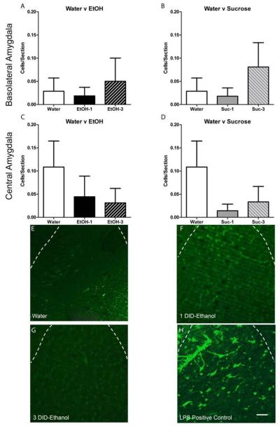
Figure 3.
Neither ethanol (A,C) nor sucrose (B,D) induced any changes in FJC+ cells. Representative photomicrographs of the FJC stain for mice that received water (E) or underwent 1 (F) or 3 (G) DID cycles of or ethanol show very little FJC+ cells. An LPS treated positive control can be seen in panel H. Scale bar in panel H= 20μm; dashed lines delineate the external capsule surrounding the basolateral amygdala. All data are presented as mean ± SEM.

