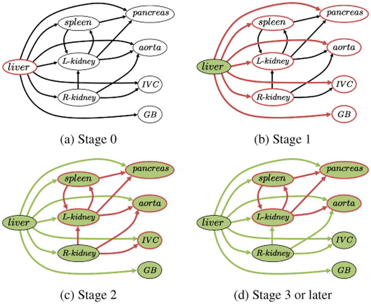
Figure 9.

Processes of multi-stage updating in multi-organ segmentation based on OCG. Red contoured nodes indicate those to be segmented. Green-colored nodes indicate those that have already been segmented. (a) Stage 0. The liver (only one organ in V1) is firstly segmented. (b) Stage 1. The organs connected from the sub-shapes of the liver are segmented. (c) Stage 2. The organs connected from those segmented at the previous stage are segmented. Note that the R-kidney and GB are not segmented at this stage because the segmentation results of predictors (only the liver in this case) do not change. (d) Stage 3 or later. Further segmentations are performed using updated results of the predictors.
