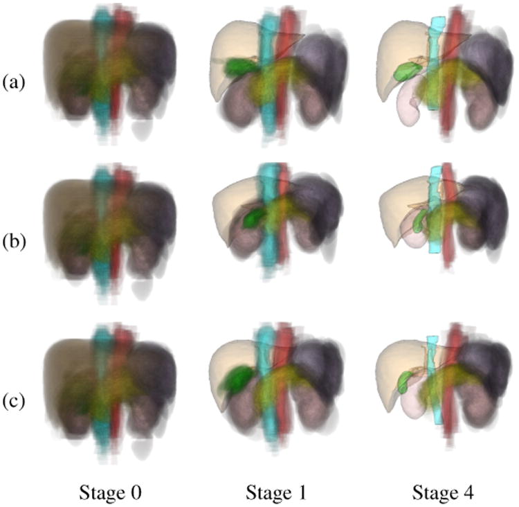
Figure 10.

Examples of prediction-based PAs at different stages of multi-organ segmentation. (a) Case 1. (b) Case 2. (c) Case 3. Left: Conventional PA of eight organs (Stage 0). Middle: Prediction-based PAs using the segmented liver (Stage 1). Right: Prediction-based PAs using the segmented liver, spleen, and kidneys (Stage 4). Orange indicates the liver, purple the spleen, pink the kidneys, yellow the pancreas, green the GB, red the aorta, and cyan the IVC. PAs are illustrated by cloud-like semi-transparent volume rendering. The segmented organ shapes are represented by semi-transparent surface rendering. At Stage 4, segmentation of R-kidney, gallbladder, and IVC (in addition to the liver) stops updating, and thus their segmented surfaces are shown instead of PAs.
