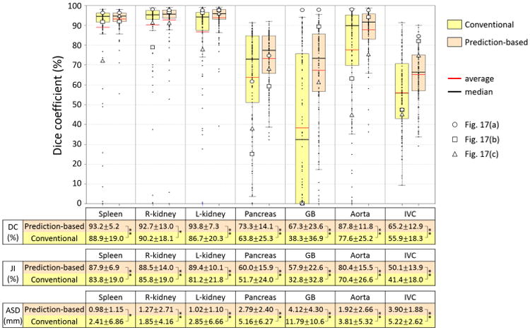
Figure 12.

Summary of accuracy evaluation for proposed prediction-based and conventional shape–location priors when unsupervised IC-IM was used for intensity model. Dice coefficients (DCs) of 134 CT data using prediction-based and conventional shape–location priors for each of seven organs were plotted. The averages of DC, Jaccard index (JI), and average symmetric surface distance (ASD) as well as statistical significance are also shown in a table format. In the table, * and ** indicate that significant accuracy improvement with significance levels of 0.05 and 0.01 was observed, respectively. DCs of three cases indicated in Figs. 17(a), (b), and (c) are plotted by ○,□, and Δ, respectively.
