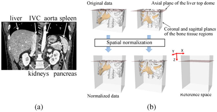
Figure 1.

Spatial normalization. (a) Coronal view of typical our CT data. (b) Definition of the abdominal normalized space. In spatial normalization, the xyz-translation and xy-scaling are adjusted so that the reference planes are aligned among all the CT data. The z-scaling is not performed because stable image features were not available for the z-scale determination. In addition, strong correlation was not found between the xy- and z-scaling at least for the liver shape. Although only sagittal planes are shown in the figures, coronal planes are similarly defined. See Appendix A for an automated method for the spatial normalization.
