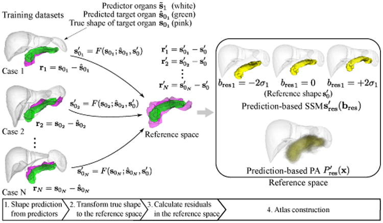
Figure 3.

Construction of prediction-based shape–location priors in reference space. Green shapes in training dataset and reference space indicate the predicted and reference shapes of the target organ (which is the pancreas in this figure), respectively. Pink shapes in training dataset and reference space indicate the true shapes and transformed true shapes of the target organ, respectively. White shapes indicate the predictor organs, which are the liver and spleen in this case. After predicting the target organ shape from predictor organs, true shapes are transformed to reference space. Then SSM and PA of the residuals are constructed in the reference space.
