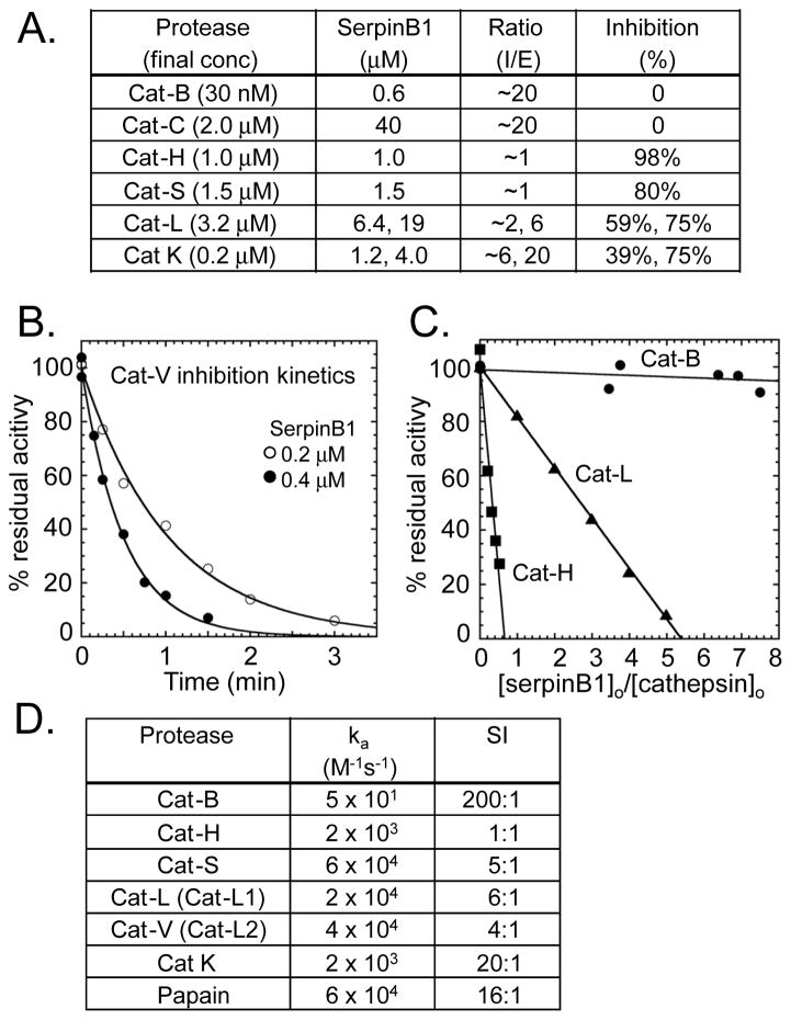
Fig. 3.
Inhibition of cathepsins by SerpinB1. A) Screening assay. Pure cathepsin and SerpinB1 were co-incubated for 5 min at near neutral pH and residual activity was assayed. I:E (inhibitor: enzyme) ratios are approximate because enzyme concentration was based on suppliers’ information. B–D) Kinetics and stoichiometry of SerpinB1 inhibition of recombinant human cathepsins at pH 6.0. B) Kinetics. Progress curves for inhibition of catV (0.5 nM) by SerpinB1. C) Stoichiometry of inhibition (SI) for catH, catL and catB by the indicated molar ratios of SerpinB1. D) Apparent second-order rate constants and SI for SerpinB1 inhibition of cathepsins. (B and C) Representative data and (A and D) average results of 2–3 experiments.

