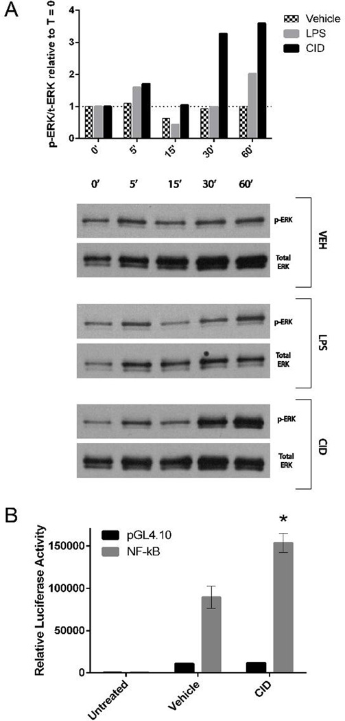
Figure 6. CID-treated MΦ-cTLR4 Cells Activate the MyD88-dependent Pathway.
(A) Western blot probing for p-ERK and total ERK. Cells were lysed at the time points indicated following CID drug, LPS, or vehicle treatment. Top panel shows phosphorylated over total ERK ratio relative to the zero timepoint for each subsequent timepont. Bottom panel shows corresponding blots (B) A Dual-luciferase® assay was used to determine NF-κB activity in MΦ-cTLR4 cells. Luciferase activity was measured 4 hours following treatment of MΦ-TLR4 cells with CID drug, LPS, or vehicle treatment. Luciferase activity was normalized to baseline Renilla luciferase activity.

