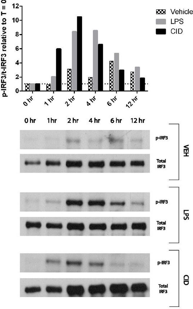
Figure 7. CID-treated MΦ-cTLR4 Cells Activate the MyD88-independent Pathway.
Western Blot for p-IRF3 and total IRF3. Cells were lysed at timepoints indicated following CID drug (50 nM), LPS (100 ng/mL), or vehicle treatment. Top panel shows phosphorylated over total IRF3 ratio relative to the zero timepoint for each subsequent timepoint. Bottom panel shows corresponding blots.

