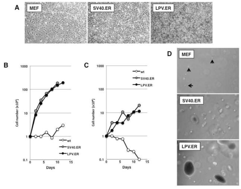
Figure 1. LPV.ER expression leads to enhanced growth and transformation of MEFs.
A) Equal amounts of wild-type MEFs, MEFs expressing SV40.ER or MEFs expressing LPV.ER were plated in cell culture and allowed to proliferate. Images (original magnification: 50×) were taken 2–3 days after plating. B) Growth curve analysis of MEFs expressing SV40 or LPV.ER. Wild-type MEFs or MEFs expressing the SV40.ER or LPV.ER were plated in 6 cm dishes and counted a various times as described in materials and methods. C) Growth curve analysis in media containing 1% serum. Wild-type MEFs or MEFs expressing the SV40 or LPV ER were plated in 6 cm dishes and grown in media supplemented with 1% FBS as described in materials and methods. Cells were counted at various times after plating. D) Anchorage independent growth of wild type, SV40.ER and LPV.ER-expressing MEFs. Cell were suspended in media containing agar supplemented with 10% FBS (materials and methods), and monitored for 3–4 weeks until colonies developed. Images were taken at the same time for all cultures at same magnification.

