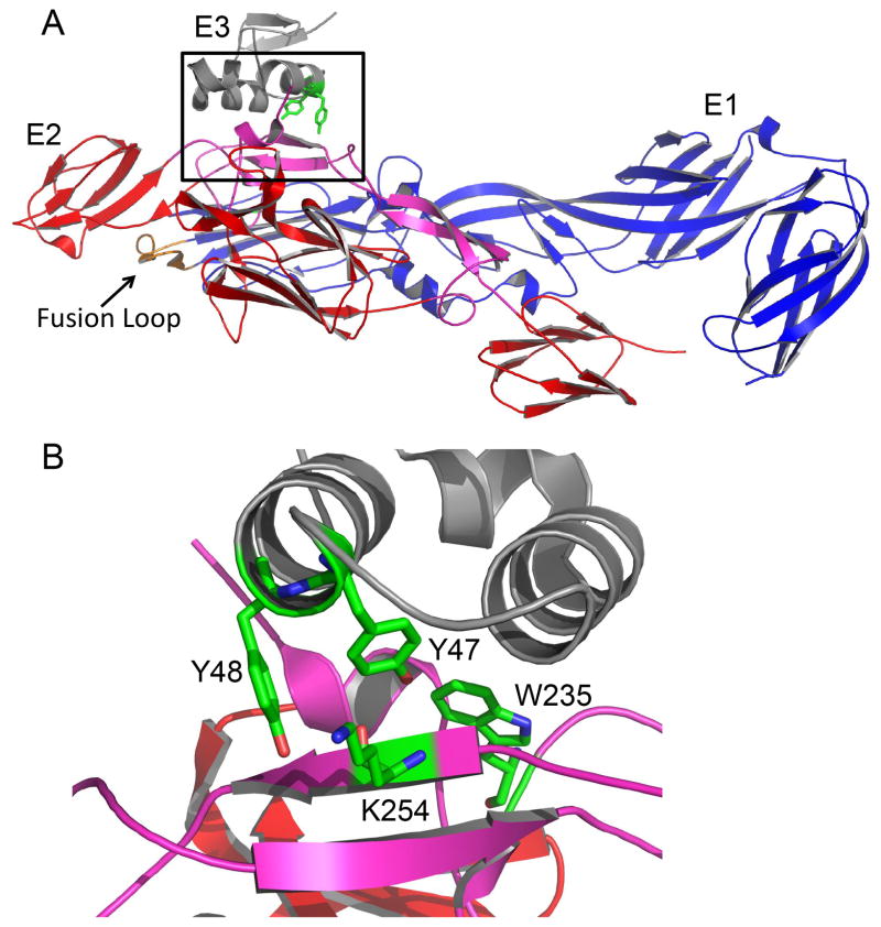
Figure 1. Alphavirus envelope protein structure and interactions at the E3-E2 interface.
A. The structure of the CHIKV E3-E2-E1 complex. E3 is shown in grey, E2 is in red with the β-ribbon connector region in purple, and E1 is in blue with the fusion loop in orange (Protein Data Bank accession number 3N42) (Voss et al., 2010). The E3-E2 interface is highlighted by the black box. B. A close up view of the E3-E2 interface (rotated from panel A) showing E3 Y47 and Y48 and E2 K254 and W235 in green stick view. Colors are as in panel A. Figure was prepared using the program PYMOL (DeLano, 2002).

