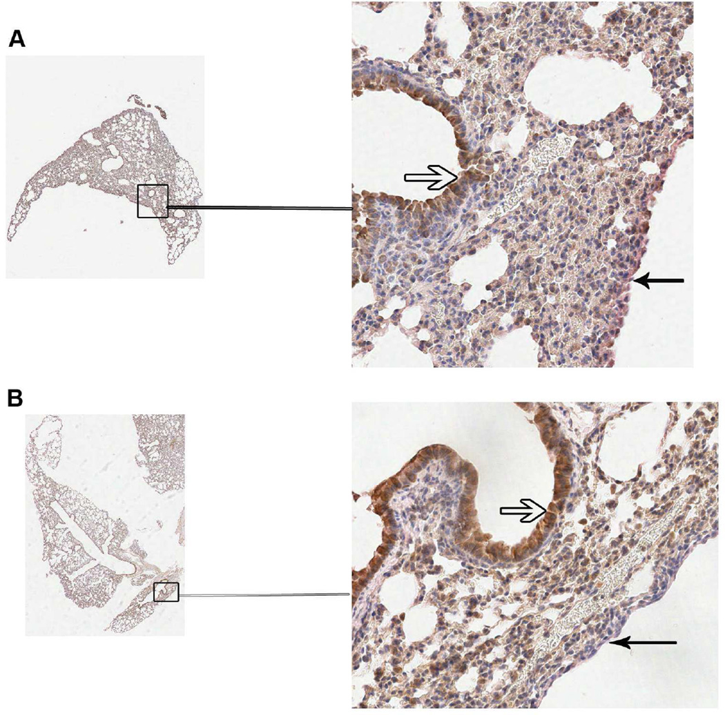
Fig. 6.
IL-33 expression in the lung is not altered by L. sigmodontis infection. Immunohistochemical staining for IL-33 in formalin-fixed lung tissue from 3 control and 3 L. sigmodontis-infected mice from day 36 and day 42 post-infection was performed. Pictured are representative sections from a control (A) and infected (B) lung at day 42. The open arrows denote bronchial epithelial cells and closed arrows indicate the lung pleural epithelium.

