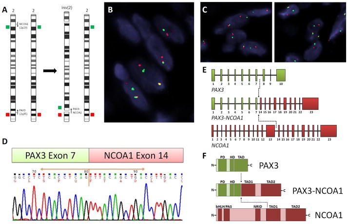
Figure 2. Novel PAX3-NCOA1 fusion in BSNS.
Schematic diagram of inv(2)(p23q35) and the corresponding FISH probes applied, flanking the PAX3 and NCOA1 genes (arrows indicate the direction of transcription)(A). FISH fusion assay showed the PAX3 (red) and NCOA1 (green) signals joined together (B), as well as the individual break-apart assays showing PAX3 (C, left) and NCOA1 (C, right) rearrangements in case #1 (red, centromeric; green, telomeric). RT-PCR chromatogram of case #2 demonstrates exon 7 of PAX3 gene joined to exon 14 of NCOA1 gene (D), resulting in a PAX3 ex1-7 - NCOA1 ex14-23 chimeric transcript (E). The predicted fusion protein retained the DNA-binding domain (DBD), including PD and HD of PAX3, and the transcriptional activation domain (TAD) of NCOA1 (F). PD, paired domain; HD, homeodomain; bHLH/PAS, basic helix-loop-helix-Per/Ah receptor nuclear translocation/Sim motif; NRID, nuclear receptor interacting domain.

