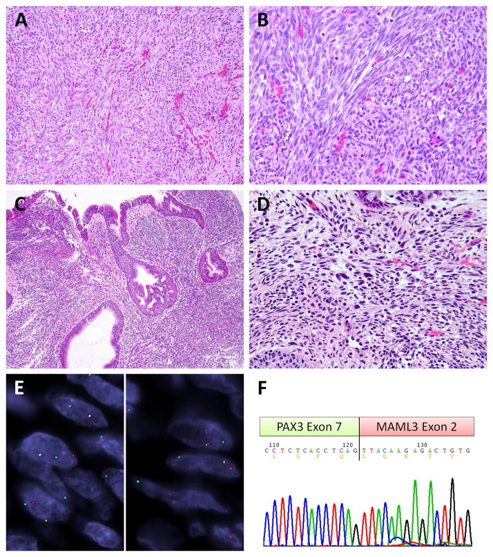
Figure 3. PAX3-MAML3 fusion positive BSNS.
All three cases displayed similar histology with sweeping or intersecting bundles (A) of monotonous and mitotically inert spindle cells (B) as exemplified by case #3. Case #5 showed respiratory type glandular hyperplasia and invaginations, mimicking inverted papilloma (C), and had scattered mast cells and rare pleomorphic hyperchromatic cells (D). The canonical PAX3-MAML3 fusion is illustrated by the break-apart signals of PAX3 (E, left) and MAML3 (E, right) by FISH in case #3 (red, centromeric; green, telomeric). The RT-PCR chromatogram of case #4 confirmed the classic fusion variant of PAX3 exon 7 to MAML3 exon 2 (F).

