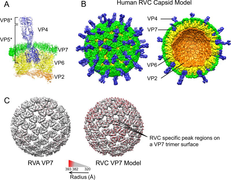
Figure 1.
Human RVC capsid model. (A) RVC 31-mer asymmetric unit model consisting of 3 VP4 (blue), 13 VP7 (green), 13 VP6 (yellow) and 2 VP2 (orange) proteins. VP4 head (VP8*) and body/stalk/foot (VP5*) illustrate products of trypsin cleavage. (B) Intact RVC icosahedral capsid model formed from the asymmetrical unit (left) and a cross-section of the capsid model (right). (C) RVA and RVC model VP7 layers colored according to radius from the center using Chimera. In order to emphasize the difference between the two VP7 layers, radii below 382 Å are shown in gray.

