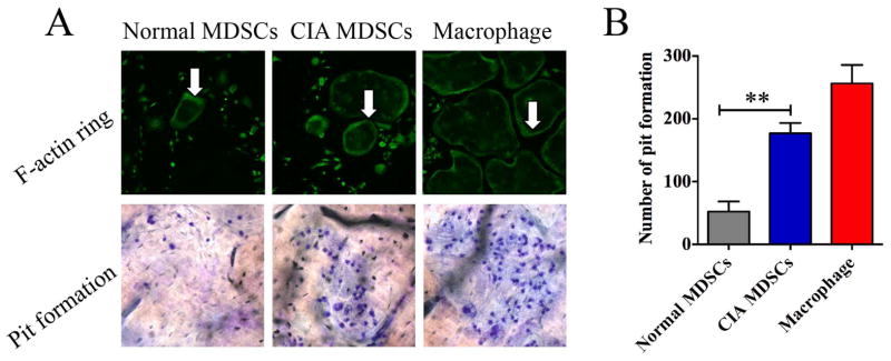
Figure 3. Bone resorption by osteoclasts derived from MDSC isolated from mice with CIA and bone marrow derived macrophages.
(A) MDSCs isolated from mice with CIA or normal mice were cultured in the presence of 50ng/ml M-CSF and 100ng/ml RANKL for 12 days. Cells were fixed and stained with FITC-phalloidin (white arrows). For the bone resorption analysis, cells were cultured with bovine cortical slides layered at the bottom of culture plate for 15 days and the cortical slides were stained with toluidine blue. Representative images are presented. (B) Pit formation on each slide was counted. Bone marrow derived macrophages were used as positive controls. Data are shown as mean ± SD. The results represent three independent experiments. **p <0.01.

