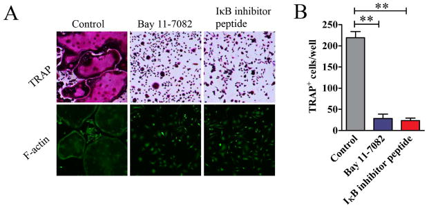
Figure 5. NF-κB signaling pathway in the differentiation of osteoclast from MDSCs.
MDSCs from the bone marrow of mice with CIA or normal mice were cultured in the presence of 50ng/ml M-CSF and 100ng/ml RANKL with or without Bay 11-7082 or IκB kinase inhibitor peptide for 12 days. (A) Cells were stained for TRAP or with FITC-phalloidin. Representative images are presented. (B) TRAP+ osteoclasts in each well were quantified and are shown as mean ± SD. The results represent three independent experiments. **p<0.01

