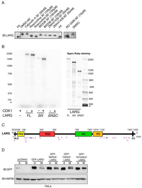
Figure 3. LARG is phosphorylated by Cdk1 during mitosis.
(A) Inhibition of Cdk1 abolishes the slower mobility of mitotic LARG. Mitotic cells, arrested after nocodazole treatment, were collected and re-plated on poly-L-lysine in the continued presence of nocodazole and in presence of the indicated mitotic kinase inhibitors or Calyculin A or vehicle control (DMSO) for 30 or 60 minutes. Cell lysates were immunoblotted using anti-LARG antibody. (B) Full-length LARG and ΔN LARG are phosphorylated in vitro by Cdk1. Purified proteins (1 μM) from Sf9-baculovirus system (right panel) were incubated with CDK1-cyclin B (13 nM) in incubation buffer (containing 20 μM [gamma-32P]ATP) for 5 min at 30°C. Samples were analyzed by SDS-PAGE followed by autoradiography (left panel). (C) Domain structure of LARG with potential Cdk1 Sites. Amino acid numbering is indicated in the above figure. Domains: PDZ, post synaptic density 95/disc-large/zona occludens; RGS, regulator of G protein signalling; DH, Dbl homology; PH, pleckstin homology; NLS, nuclear localization signal; CC, coiled-coil. The pink stars indicate potential Cdk1 sites. The dark blue stars indicate the sites that are the epitopes targeted by the pS190 and pS1176 phosphospecific antibodies. (D) Mitotic-dependent gel mobility shift of LARG is altered by N- and C-terminal phosphonull mutations. HEK293 cells were transfected with the indicated constructs, treated with nocodazole or vehicle control. Nocodazole-treated cells were subjected to mitotic shake-off, and cell lysates were prepared (M). Cell lysates were also prepared from asynchronously growing cells (A). Lysates were then immunoblotted using an anti-GFP antibody.

