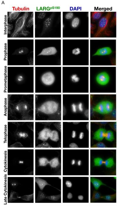
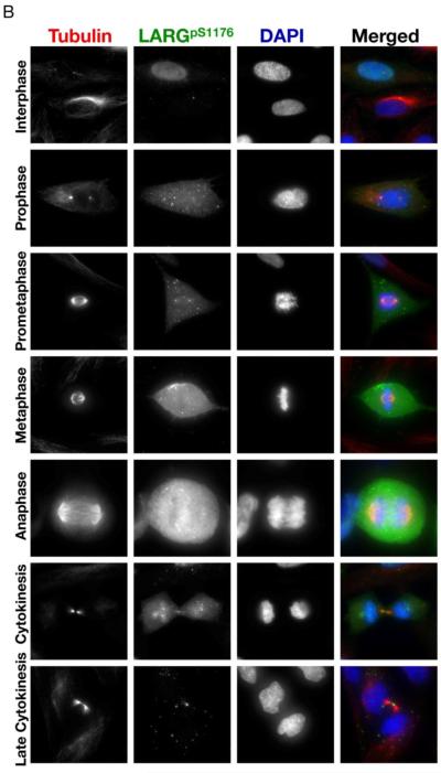
Figure 5. LARG phosphorylated at sites S190 and S1176 localizes to the centrosomes and flanks the midbody in cytokinesis.
(A) HeLa cells were fixed and stained with anti-α-tubulin antibody (red), anti-pS190 LARG (green), and DNA was visualized by DAPI staining (blue). (B) Same as in left panel, except anti-pS1176 LARG is in green. Cells in different stages of mitosis were identified based on hallmark nuclear and microtubule staining.


