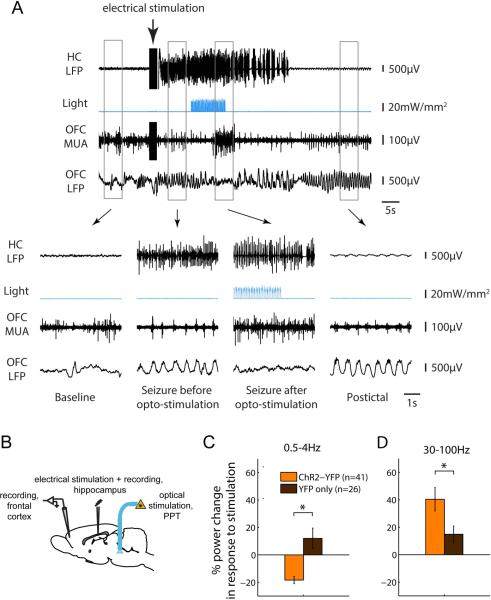
Figure 2. Optogenetic stimulation of the PPT reduces cortical slow wave activity during seizures.
We induced focal seizures by injecting a brief (2sec) electric current via a bipolar electrode implanted into the hippocampus (see part B for schematic). The same electrode was used for recording hippocampal local field potential (LFP). 10s after seizure onset, we delivered a 10s-long train of blue light pulses (473nm; pulse duration 40ms, frequency 10Hz) into the PPT. We also recorded LFP and multiunit activity from the orbitofrontal cortex (OFC). In control seizures (not shown), in which YFP but no ChR2 had been injected into the PPT, cortical slow wave activity persisted throughout the seizure as previously described 8; 9. (A) Optogenetic stimulation of the PPT following ChR2 injection resulted in a transition of the cortical LFP from slow waves to low voltage fast activity. (B) Schematic of seizure experimental setup. (C-D) Changes in cortical LFP delta (0.5-4Hz) and gamma (30-100Hz) power in response to optogenetic stimulation. LFP Power was quantified shortly after seizure onset and following optogenetic stimulation (5-15s and 18-28s after seizure onset, respectively), and the percent change in power between the two time windows was averaged across seizures. * P<0.05.

