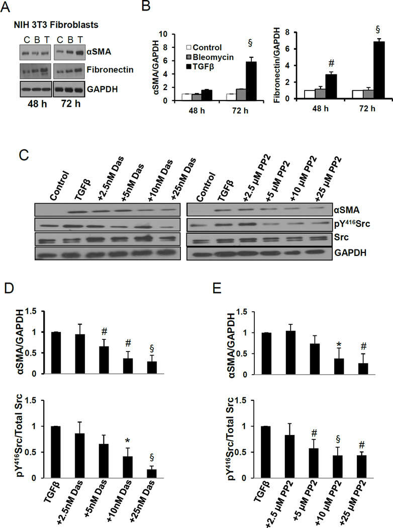
Fig. 1.
Dasatinib inhibits TGFβ-induced myofibroblast differentiation and αSMA expression. A. Western blot images of NIH 3T3 lysates treated in the presence and absence of 100 pM TGFβ or 2.5 mU bleomycin for 48, and 72 h, probed for αSMA and fibronectin. B. Densitometry analysis of the Western bands showing expression changes in αSMA and fibronectin with 100 pM TGFβ and 2.5 mU bleomycin for 48, and 72 h and normalized to GAPDH (n=3–6). C. Western blot images of NIH 3T3 lysates treated in the presence and absence of 100 pM TGFβ (48 h) and in combination of TGFβ (48 h total) with various doses of dasatinib or Src inhibitor PP2 (24 h), probed for αSMA, pY416Src and total Src. D. Densitometry analysis of the Western bands showing expression changes in αSMA and pY416Src with 100 pM TGFβ combined with various doses of dasatinib, and normalized to GAPDH and total Src, respectively. (n=3). E. Densitometry analysis of the Western bands showing expression changes in αSMA and pY416Src with 100 pM TGFβ combined with various doses of Src inhibitor PP2 and normalized to GAPDH and total Src, respectively (n=3). Data presented as mean ± S.D. *P<0.05; #P<0.01; §P<0.001

