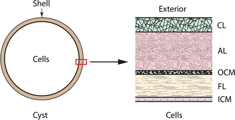
Fig. 2.
Schematic representation of the Artemia cyst shell. A cyst in cross section is shown in the left side of the diagram. The right side of the figure, showing the ultrastructure of the cyst shell, is an enlargement of the portion of the shell enclosed in the red box. CL cuticular layer, AL alveolar layer, OCM outer cuticular membrane, FL fibrous layer, ICM inner cuticular layer. The relative widths of the cyst shell layers shown in the figure approximate the relative widths of the layers seen in samples prepared for electron microscopy. The figure was adapted from Drinkwater and Clegg (1991)

