
Figure 1.
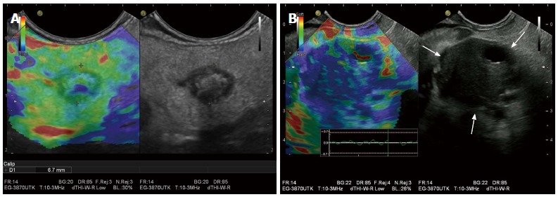
Benign and malignant pancreatic masses on endoscopic ultrasound elastography. A: A pancreatic teratoma is shown as heterogeneous soft (green) pattern (left: EUS elastography image; right: B-mode image); B: A pancreatic ductal adenocarcinoma appears stiffer (blue) than the adjacent normal pancreatic parenchyma, probably due to the presence of fibrosis and marked desmoplasia.