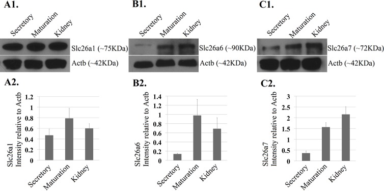
Fig 3. Western blot analysis of Slc26a1, Slc26a6 and Slc26a7.
A1-C1. Protein-level expression of Slc26a1, Slc26a6 and Slc26a7 was detected by western blot analysis using samples obtained from both secretory- and maturation-stage enamel organs (4-week-old rat incisors). Protein samples extracted from kidney (4-week-old rat) were used as reference controls. The molecular weights for Slc26a1, Slc26a6 and Slc26a7 are 75kDa, 90kDa and 72kDa, respectively. Beta-Actin served as the control for sample loading. A2-C2. The intensities of the bands (relative to Beta-Actin) were measured using ImageJ. The average fold changes of Slc26a1, Slc26a6 and Slc26a7 at the protein level were ~1.6, ~6.1, ~4.2, respectively.

