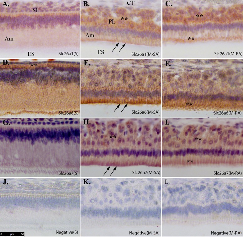
Fig 4. Immunoperoxidase immunostaining of Slc26a1, Slc26a6 and Slc26a7 in secretory- and maturation-stage enamel organ.
Immunostaining procedures were applied to the sagittal sections prepared from paraffin-embedded 4-week-old rat mandibles. A. Slc26a1 in secretory-stage ameloblasts (S); B. Slc26a1 in smooth-ended ameloblasts at maturation stage (M-SA); C. Slc26a1 in ruffle-ended ameloblasts at maturation stage (M-RA); D. Slc26a6 in secretory-stage ameloblasts (S); E. Slc26a6 in smooth-ended ameloblasts at maturation stage (M-SA); F. Slc26a6 in ruffle-ended ameloblasts at maturation stage (M-RA); G. Slc26a7 in secretory-stage ameloblasts (S); H. Slc26a7 in smooth-ended ameloblasts at maturation stage (M-SA); I. Slc26a7 in ruffle-ended ameloblasts at maturation stage (M-RA); J-L. The sections that were incubated without antibodies served as negative controls for immunostaining. All images were collected under 20x magnification. Scale bar shown in Panel J (50μm). Slc26a1, Slc26a6 and Slc26a7 all showed expression on the apical membrane and/or within subapical cytoplasmic region (double black arrows). Positive staining in other regions was indicated by double black asterisks. SI—Stratum intermedium; Am—Ameloblast; ES—Enamel space; CT—Connective tissue; PL—Papillary layer.

