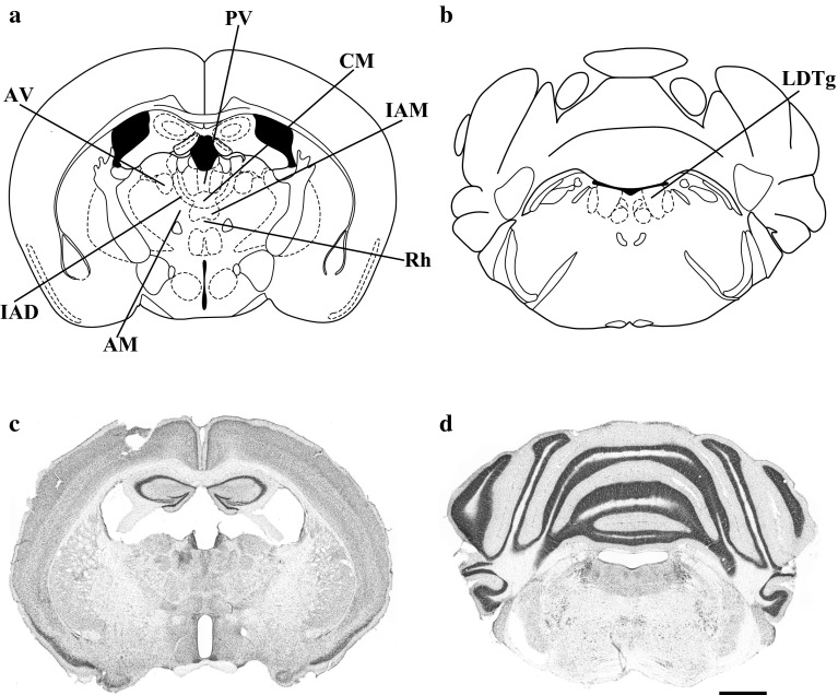
Fig. 2.
Line drawing presenting a the thalamic nuclei and b laterodorsal tegmental nuclei, which reveal the highest level of β-galactosidase expression in B6.Cg-Tg(Nes-cre)1Nogu mice. c, d Cresyl violet-stained coronal section of B6.Cg-Tg(Nes-cre)1Nogu mouse brain at the same level as in (a, b). AM anteromedial thalamic nucleus, AV anteroventral thalamic nucleus, CM central medial thalamic nucleus, IAD interanterodorsal thalamic nucleus, IAM interanteromedial thalamic nucleus, PV paraventricular thalamic nucleus, Rh rhomboid thalamic nucleus, LDTg laterodorsal tegmental nucleus. Scale bar 1 mm

