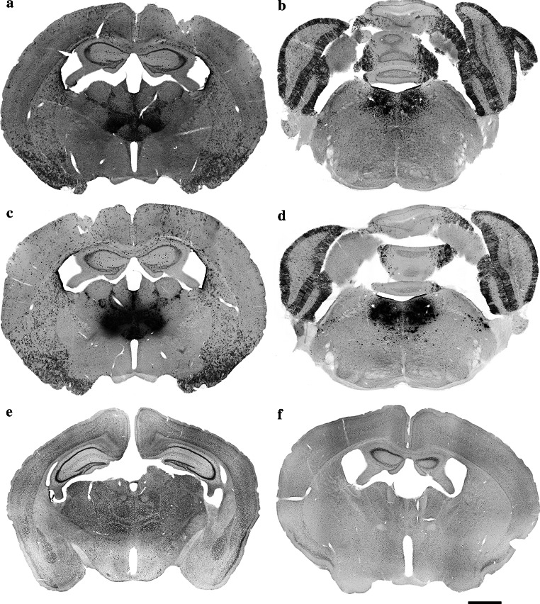
Fig. 5.
Low-magnification images showing the expression of β-galactosidase in the thalamic nuclei (a, c) and laterodorsal tegmental nuclei (b, d) of the B6.Cg-Tg(Nes-cre)1Nogu mouse brain, revealed by a combination of X-gal (a, b) or Salmon-gal (c, d) and tetranitroblue tetrazolium. Reaction time is 24 h for the X-gal/tetranitroblue tetrazolium and 5 h for Salmon-gal/tetranitroblue tetrazolium. Control sections from wild-type mice at the level of the thalamus stained with X-gal (e) or Salmon-gal (f) and tetranitroblue tetrazolium. Scale bar 1 mm

