Figure 7.
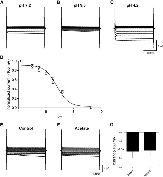
CLH-1 is sensitive to extracellular pH but insensitive to intracellular pH. A–C, CLH-1 currents in an oocyte that was perfused with solutions at pH 7.2 (A), pH 9.3 (B), and pH 4.2 (C), using the same voltage protocol as in Figure 6. D, Sigmoidal dose–response curve of currents from CLH-1-expressing oocytes (n = 7) at −160 mV, normalized to pH 4.2 as maximum and pH 9.3 as minimum activation. LogEC50 = pH 6.778 ± 0.1050 for extracellular pH activation of CLH-1. Data are mean ± SEM. E, F, Example traces of currents from CLH-1-expressing oocytes that had been preincubated for 1–2 h in control buffer (E) or 20 mm acetate (F) clamped at voltages ranging from −160 mV to 100 mV. G, There was no significant difference in CLH-1 currents between control (−1.10 ± 0.40 μA, n = 3) or acetate-incubated (−1.04 ± 0.35 μA, n = 4) oocytes at a voltage of −160 mV. Data are mean ± SEM. p = 0.9154 (t test).

