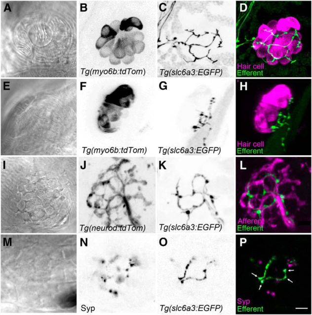
Figure 3.
Dopaminergic fibers are located beneath the hair-cell layer in neuromasts. A–D, Top-down view of a neuromast. Hair cells are stably expressing tdTomato (magenta), whereas DA efferent fibers express GFP (green). E–H, Side view of a neuromast. Efferent fibers are closely positioned near the basal ends of hair cells but do not appear to make direct synapses. GFP-positive varicosities appear randomly with respect to hair-cell position. I–L, Top-down view of the extensive pattern of afferent fiber innervation (magenta) in contrast to the relatively sparse innervation by DA efferents (green). DA fibers track in parallel with a subset of afferent fibers. M–P, Immunolabel of the synaptophysin (Syp) synaptic vesicle marker indicates vesicles within the varicosities of the GFP-positive fibers (arrows) and also indicates clusters of vesicles within other types of efferents. Note the absence of juxtaposition of non-DA efferent vesicles (magenta only) and dopaminergic fibers. B–D, J–L, Maximum projections. F–H, 3D reconstructions of maximum projections. A, E, I, M–P, Single optical slices. Scale bars: A–P, 5 μm.

