Figure 4.
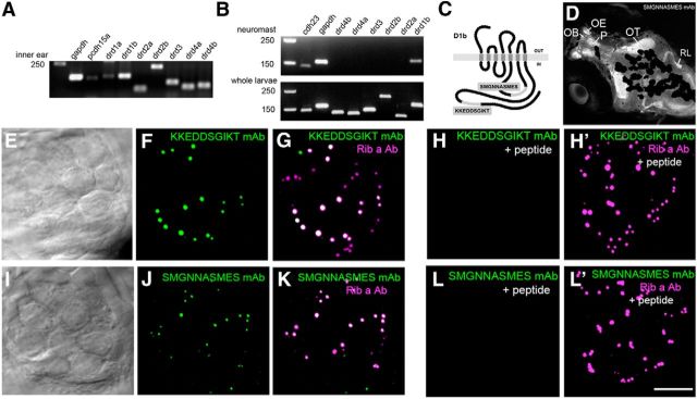
Expression of DA receptor subtypes in zebrafish. A, RT-PCR of drd genes from adult inner ear cDNA that includes both epithelial and neuronal cell types. B, RT-PCR of drd genes in neuromast and whole larval cDNA. DNA markers are present in the first lane, followed by positive controls (gapdh and pcdh15a or cdh23). C, Diagram of D1b protein depicting the location of the amino acid sequences used to generate monoclonal antibodies. D, Immunolabel (SMGNNASMES mAb) of the head of a 5 dpf larva. The focal plane includes the dorsal region of the fish head, and signals are prominent within the forebrain, tectal, and hindbrain regions. Specific regions are indicated. Melanocytes partially occlude the labeling in deeper medial areas of the brain. OB, Olfactory bulb; OE, olfactory epithelium; OT, optic tectum; P, pallium; RL, rhombic lip. E–F, I–J, Immunolabel of neuromast epithelia with D1b mAbs. DIC image (single optical plane) and corresponding immunofluorescence for KKEDDSGIKT (E, F) and SMGNNASMES mAbs (I, J; maximum projections are shown). Colocalization with Ribeye a is shown in G, K. H, H′, L, L′, Peptide blocking experiments with KKEDDSGIKT and SMGNNASMES peptides using the corresponding D1b antibodies. Ribeye a antibody was included as a control in H′, L′. Scale bars: D, 75 μm; E–L′, 5 μm.

