FIGURE 7.
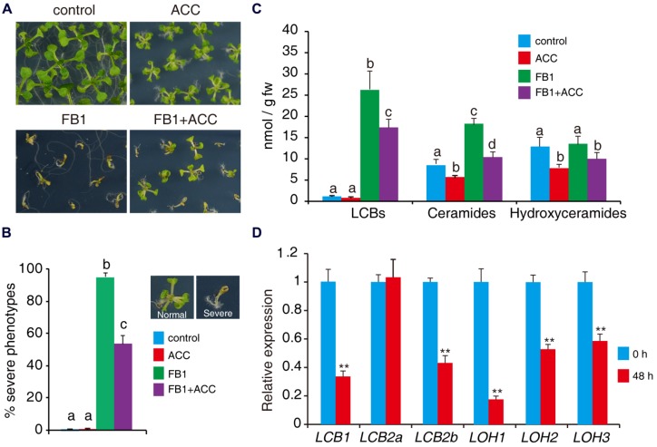
ACC reduces FB1-induced cell death. (A) Wild-type seeds were germinated on 1/2x MS supplemented with various combinations of 0.5 μM FB1 and 50 μM ACC. Photos were taken after 2 weeks of treatment. (B) Quantitative analysis of the phenotype of seedlings in (A). At least 100 seeds of each sample were used in each experiment. Data presented are means ± SD (n = 3) and sets marked with different letters indicate significances assessed by Student–Newman–Keul test (P < 0.05). (C) Sphingolipid analysis of wild-type seedlings treated with various combinations of ACC and FB1. One-week-old seedlings were used and harvested after 6 days of treatment. Total sphingolipids were extracted and analyzed (see Supplementary Figure S2 for the detailed analysis). (D) Expression of genes involved in de novo sphingolipid synthesis in wild-type seedlings after 48 h ACC treatment. Data represent means ± SD from three technical repeats and sets marked with double asterisks indicate significance assessed by Student’s t-test (P < 0.01). This experiment was repeated three times with similar results.

