
FIGURE 3.
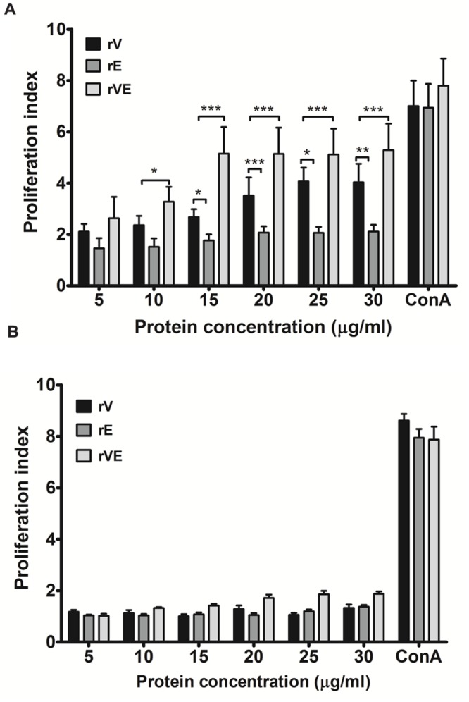
Proliferation assay performed with lymphocytes isolated from the spleen of immunized and sham immunized groups of mice at 43rd day. (A) Lymphocytes isolated from the rV, rE, and rVE immunized mice were cultured in 96-well flat bottom plates with a gradient concentration of purified recombinant proteins. After 72 h of incubation at 37°C (5% CO2), proliferative responses were determined by MTT assay. (B) Proliferation assay performed with lymphocytes obtained from sham immunized mice spleen and were stimulated with gradient concentration of purified recombinant proteins (rV, rE, and rVE). The T-cell inducer, ConA at 20 μg/ml was considered as positive control. Extracted lymphocytes with culture medium alone were used as a negative control. Data represented as PI mean values ± SD. ∗P < 0.05; ∗∗, P < 0.01; ∗∗∗, P < 0.001; compared to control group (sham immunized mice).