Figure 6.

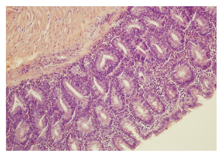
A typical microscopic image of colonic mucosa taken from the animals watered with DSS solution and treated intraperitoneally with ghrelin. Hematoxylin-eosin counterstain. Original magnification 200x.

A typical microscopic image of colonic mucosa taken from the animals watered with DSS solution and treated intraperitoneally with ghrelin. Hematoxylin-eosin counterstain. Original magnification 200x.