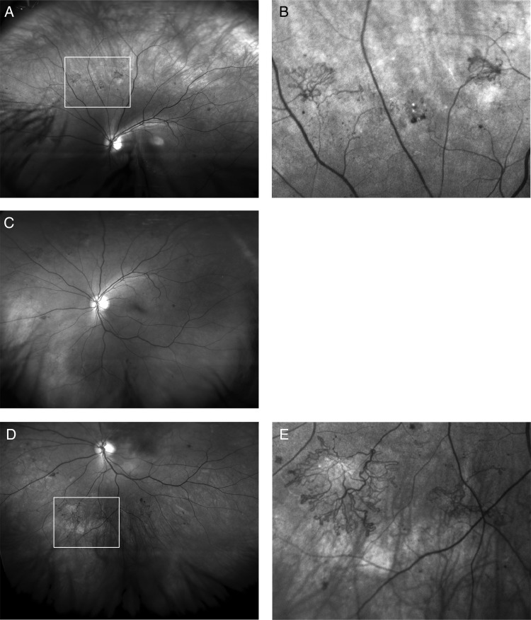
Figure 1.
Red free Optomap images of a left eye of a diabetic referred with maculopathy in the other eye, (R1, M1), the left eye being referred as, (R1, M0). (A) Up-steered, showing new vessels, (B) with zoom, (C) straight ahead showing new vessels outside two fields and on the edge of the standard seven-field images, (D) down steered, showing new vessels and (E) better seen with on zoom.

